Digestive tract abnormalities can sometimes develop with subtle changes that are difficult to identify during a standard examination. Small lesions, irregular mucosal patterns, and early tissue changes may not always be clearly visible without advanced imaging. High definition endoscopy with NBI combines detailed image resolution with specialized light wavelengths to provide gastroenterologists with a closer view of the gastrointestinal lining. This technology can help doctors identify areas that appear different from surrounding tissue, examine suspicious findings more closely, and determine whether further evaluation is needed. Understanding how this advanced imaging works can help patients appreciate its role in modern gastrointestinal diagnosis and monitoring.
High-definition endoscopy with NBI provides more detailed viewing
High-definition endoscopy uses advanced cameras and imaging systems to produce clearer, sharper views of the gastrointestinal lining. Depending on the procedure, the gastroenterologist can examine the oesophagus, stomach, duodenum, or colon in greater visual detail. Higher image resolution can make subtle differences in tissue colour, surface texture, borders, and structure easier to observe. This can be particularly useful when abnormalities are small or differ only slightly from surrounding healthy tissue. High-definition imaging therefore provides a stronger visual foundation for detailed examination. When combined with NBI, the doctor can obtain additional information about mucosal and vascular patterns. This combination allows gastroenterologists to examine the digestive tract more closely than conventional white-light imaging alone.
NBI enhances the visibility of mucosal and vascular patterns
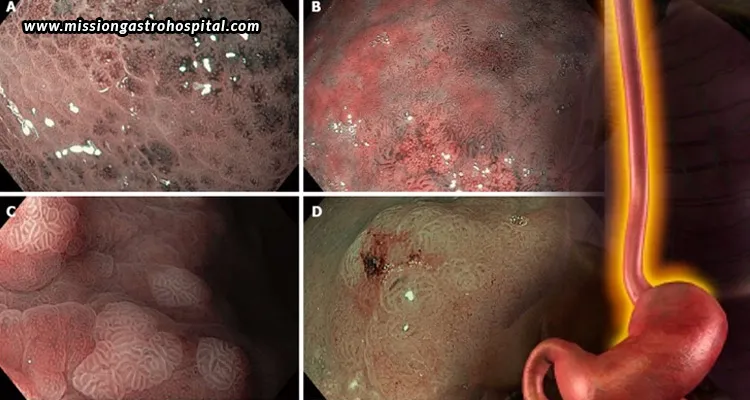
Narrow Band Imaging is an optical imaging technique designed to enhance the visibility of certain tissue and blood-vessel patterns during endoscopy. Instead of relying only on broad-spectrum white light, NBI uses selected narrow wavelengths that interact with superficial tissue and blood vessels in distinctive ways. This can increase the visual contrast between certain structures and surrounding mucosa, making surface characteristics and vascular patterns easier to observe. When an area appears unusual, NBI can provide additional visual information that helps the gastroenterologist examine it more closely. However, NBI does not independently diagnose disease or replace conventional endoscopic examination. When combined with high-definition imaging, it can support the identification and characterization of suspicious areas and contribute to a more detailed assessment of the digestive tract.
High-definition imaging helps identify subtle digestive tract abnormalities
Not every digestive tract abnormality appears as a large or clearly defined growth. Some lesions may be small, flat, slightly discoloured, or associated with subtle changes in the surrounding mucosa. These findings can require careful inspection because their appearance may differ only minimally from normal tissue. High-definition imaging provides a clearer view of these small details, allowing the endoscopist to examine the gastrointestinal lining more closely. This improved visualization can be particularly useful when the goal is to recognize abnormalities that may otherwise be difficult to distinguish. Although advanced imaging cannot guarantee that every lesion will be detected, clearer visualization can support a more thorough examination. It can also help the gastroenterologist identify areas that require closer inspection, monitoring, biopsy, or further evaluation.
NBI helps gastroenterologists examine suspicious areas more closely
When a gastroenterologist notices an area that appears different from surrounding tissue, NBI can provide a more detailed view of its surface and vascular characteristics. The endoscopist may assess features such as vascular arrangement, surface patterns, lesion borders, and overall appearance while comparing the area with nearby normal-looking mucosa. This enhanced visualization can provide useful information when a finding is difficult to characterize using conventional imaging alone. Depending on the appearance and the patient’s clinical circumstances, the doctor may determine whether the area requires monitoring, biopsy, removal, or additional investigation. NBI therefore allows the examination to move from general observation to more focused assessment. Its findings are interpreted alongside the patient’s symptoms, medical history, endoscopic appearance, and other relevant clinical information.
High-definition endoscopy with NBI supports early detection of gastrointestinal lesions
Early gastrointestinal lesions may sometimes produce few or no noticeable symptoms, making appropriate endoscopic examination important for detecting subtle abnormalities. High-definition endoscopy with NBI provides detailed views of the mucosal surface while enhancing selected vascular patterns. These enhanced images may help the endoscopist recognize areas that warrant closer examination, including subtle lesions or irregular tissue changes. Identifying a suspicious area earlier can allow the gastroenterologist to investigate it before it potentially becomes more advanced. However, NBI should not be considered a standalone cancer detection or diagnostic test. If an abnormal area is identified, the doctor may recommend biopsy or another appropriate evaluation. The final diagnosis depends on the complete clinical assessment and, when necessary, pathological examination of tissue samples.
High-definition endoscopy with NBI can help evaluate abnormalities in the oesophagus and stomach
High-definition endoscopy with NBI can be used during examinations of the upper digestive tract, including the oesophagus and stomach. In the oesophagus, enhanced imaging may help doctors examine areas with unusual mucosal appearance, irregular borders, or other changes requiring closer assessment. During stomach examinations, it can provide additional information about subtle differences in surface structure and vascular patterns. The gastroenterologist may initially examine the area using high-definition white-light imaging before applying NBI for a more detailed assessment. This combined approach can be useful when a finding is not clearly characterized during the initial inspection. Depending on its appearance and the patient’s clinical circumstances, the doctor may recommend biopsy, removal, surveillance, or further investigation.
High-definition endoscopy with NBI can improve the assessment of colon polyps and lesions
During colonoscopy, gastroenterologists examine the colon lining for polyps, abnormal tissue, and other findings. These lesions can vary in size, shape, surface appearance, and vascular characteristics. Some are easily visible, while others may be relatively subtle or flat. High-definition imaging provides a clearer view of the colonic mucosa, helping the endoscopist examine small surface details. NBI can then provide additional information about the lesion’s surface and vascular patterns, supporting closer characterization. Depending on factors such as its appearance, size, location, and clinical context, a polyp may be removed, biopsied, or monitored. Advanced imaging therefore complements careful colonoscopic examination and can support clinical decision-making, while pathological examination remains important when tissue diagnosis is required.
Advanced endoscopic imaging can help guide biopsy and further evaluation
Enhanced endoscopic imaging can help the gastroenterologist determine which areas may require further investigation. When a suspicious region is identified, high-definition endoscopy with NBI can provide a closer view of its surface and vascular characteristics. This information may help the doctor identify an appropriate area for biopsy when tissue sampling is indicated. However, advanced imaging does not replace biopsy or pathological examination when a definitive tissue diagnosis is necessary. A biopsy allows specialists to examine cells and tissue under a microscope and determine the nature of an abnormality. The endoscopic appearance, patient history, symptoms, and pathology findings may all contribute to the final diagnosis. NBI should therefore be considered a supporting tool within a broader diagnostic process rather than an independent diagnostic method.
How an experienced gastroenterologist maximizes the benefits of NBI endoscopy
Advanced imaging equipment is only one part of an effective endoscopic examination. The experience and clinical judgment of the gastroenterologist are equally important when interpreting NBI findings. An experienced endoscopist can determine when high-definition imaging is sufficient and when NBI may provide additional information. They can identify areas requiring closer inspection and decide whether biopsy, removal, surveillance, or further evaluation is appropriate. Proper preparation, careful examination, accurate documentation, and appropriate technique also contribute to procedure quality. Combining advanced technology with professional expertise can support a more comprehensive assessment of the digestive tract. Patients considering advanced gastrointestinal evaluation can consult an experienced gastroenterology team equipped to provide detailed diagnostic care. Mission Gastro Hospital in Ahmedabad offers high definition endoscopy with NBI, providing detailed visualization to support the assessment of digestive tract abnormalities and informed clinical evaluation.
High-definition endoscopy with NBI offers gastroenterologists a more detailed way to examine the digestive tract by combining sharper imaging with enhanced visualization of selected mucosal and vascular patterns. This can help identify subtle abnormalities, examine suspicious lesions more closely, and support decisions about biopsy or further evaluation. The technology can be useful across examinations of the oesophagus, stomach, and colon, although its suitability depends on each patient’s clinical circumstances. NBI does not replace pathological diagnosis or professional medical judgment. Instead, it complements conventional endoscopy and other diagnostic methods. For patients who require gastrointestinal evaluation, discussing advanced endoscopic options with an experienced gastroenterologist can help determine the most appropriate approach.